Dental X-ray film, also known as dental film, is an important means in the process of dental diagnosis and treatment. Clinical examination can only intuitively judge the condition of the crown part, while for the root, alveolar bone, periodontal ligament, etc., X-ray must be passed. sheet inspection. Teeth show white radiographic images on dental radiographs, of which enamel is the strongest, dentin and cementum are lower than enamel, the pulp cavity is black and transparent, and the root canal orifice to the apical foramen is gradually changing. thin image.
Dental tablet is the most commonly used examination method in stomatology. It is used to find the degree and scope of lesions before treatment; to guide treatment, determine the treatment range and depth of treatment during treatment; to observe the curative effect after treatment.
In addition to understanding dental lesions, X-ray films can also be used to understand whether there are impacted teeth, supernumerary teeth, odontogenic tumors and cysts in the alveolar bone. Comprehensive analysis of X-ray films can improve the accuracy and success of dental diagnosis and treatment. Rate.
The meaning and use of X-ray films
1. Maxillofacial trauma: know whether the teeth are fractured, the degree and scope of alveolar bone fractures.
2. Tooth hard tissue lesions: tooth root caries, proximal caries, determine the degree of caries and the range of caries; the relationship between caries depth and pulp angle or pulp cavity; understand caries and pulp cavity distance; understand the changes of the pulp cavity and the periapical situation.
3. Pulp lesions: dental tablets help to check pulp stone disease and internal absorption.
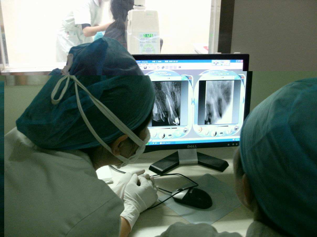
4. Periapical inflammation: understand the root canal conditions of the diseased tooth: such as the number, thickness, curvature of the root canal, and whether there is calcification and internal absorption; check the root canal filling treatment. If a tooth needs root canal treatment, three or more X-rays are required. The preoperative film is used to judge the apical inflammation, the size and extent of the cyst, etc., to judge the prognosis of root canal treatment; the intraoperative film of root canal preparation is used to check the length of root canal treatment, and the postoperative film to evaluate the success of root canal treatment. .
5. Periodontal inflammation: It shows the extent and extent of alveolar bone resorption.
6. It is used for the inspection and treatment of impacted teeth, impacted teeth and replacement teeth.
7. X-rays to determine the relative position of the deciduous and permanent teeth to help him diagnose whether the deciduous teeth can fall out on their own or need to be extracted.
8. Orthodontics: Take panoramic oral X-rays and lateral cephalograms to check the development of bones and teeth.
9. Tooth extraction Check the condition of the root and the degree of damage to the alveolar bone, whether the tooth is extracted cleanly and whether the shape of the alveolar socket is intact.
10. The dental implant film shows the health, height and density of the alveolar bone, whether it is suitable for dental implants; the survival of implant nails.
11. Size and extent of alveolar bone cysts and tumors.
12. Salivary gland stones, salivary gland angiography, temporomandibular joint.
Post time: Jul-25-2022
